Authors / metadata
DOI: 10.36205/trocar1.2022004
Abstract
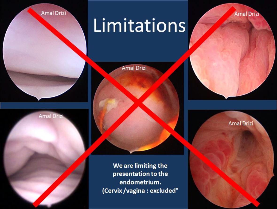
On demand of a specific entity in New Zealand a practical handout based on images backed up by histology was conceived to help the junior residents to interpret the endometrial lining of the uterine cavity. The description of the vaginal epithelium and the cervical lining are not included in the handout.
Material and Methods
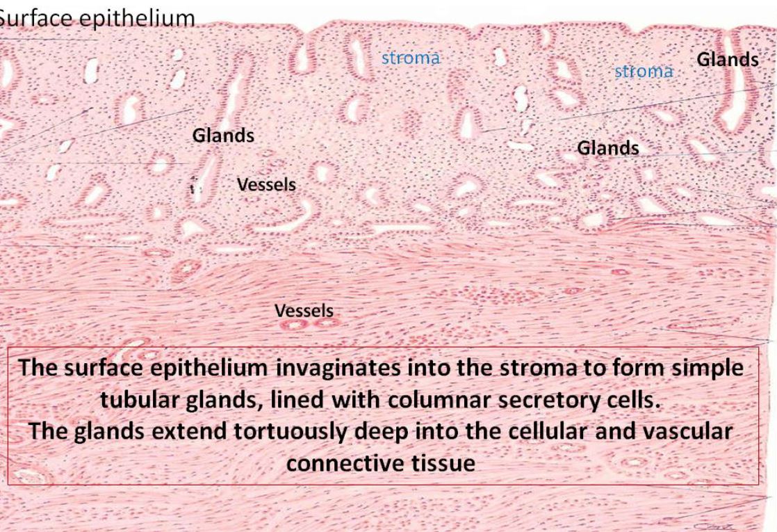
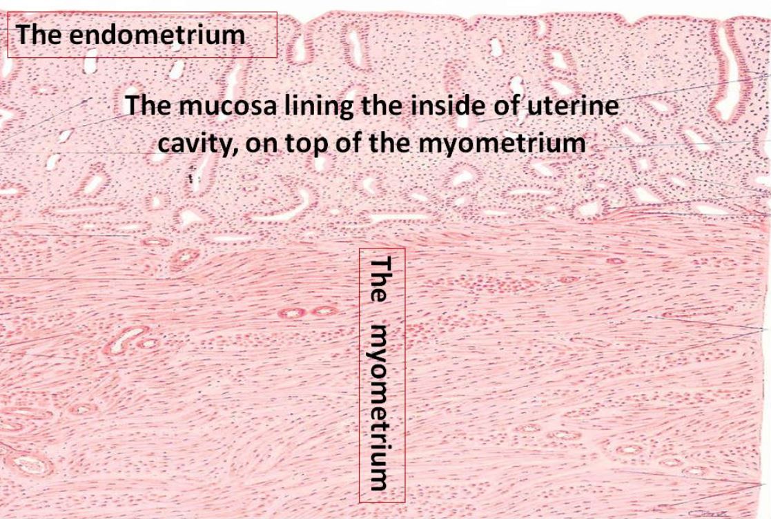
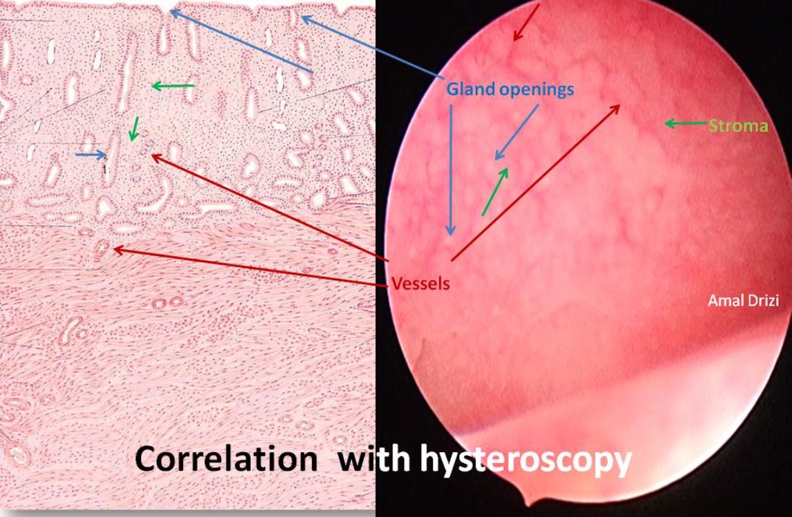
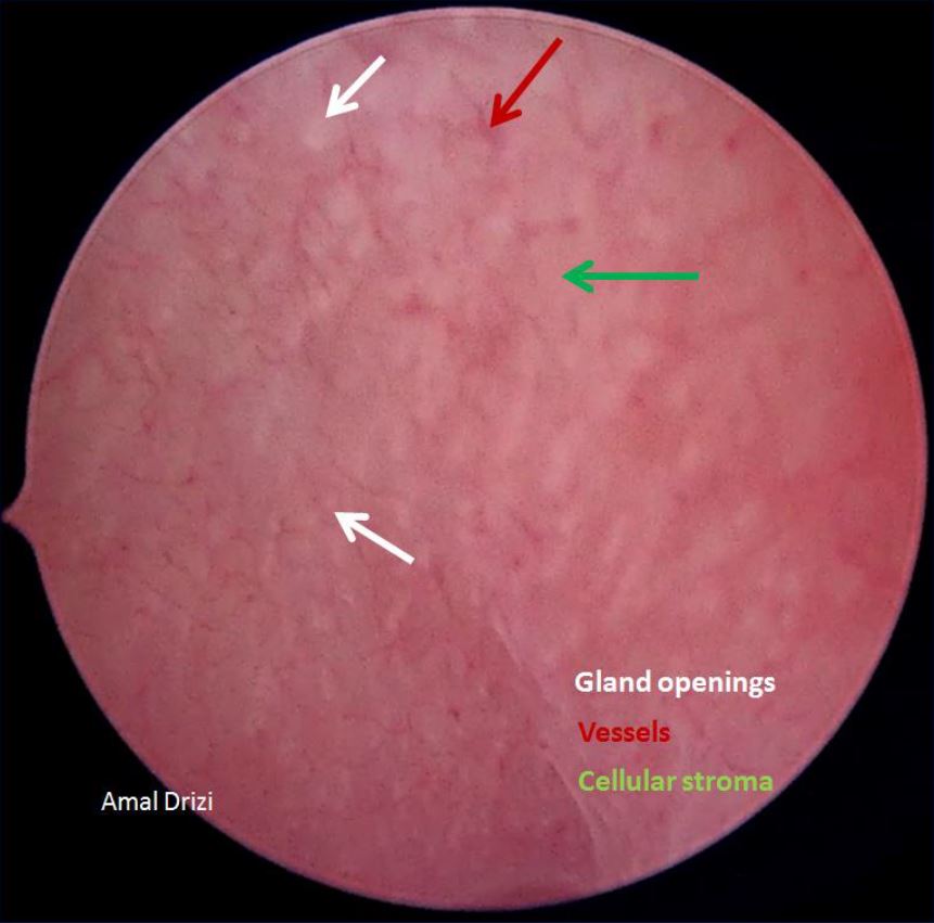
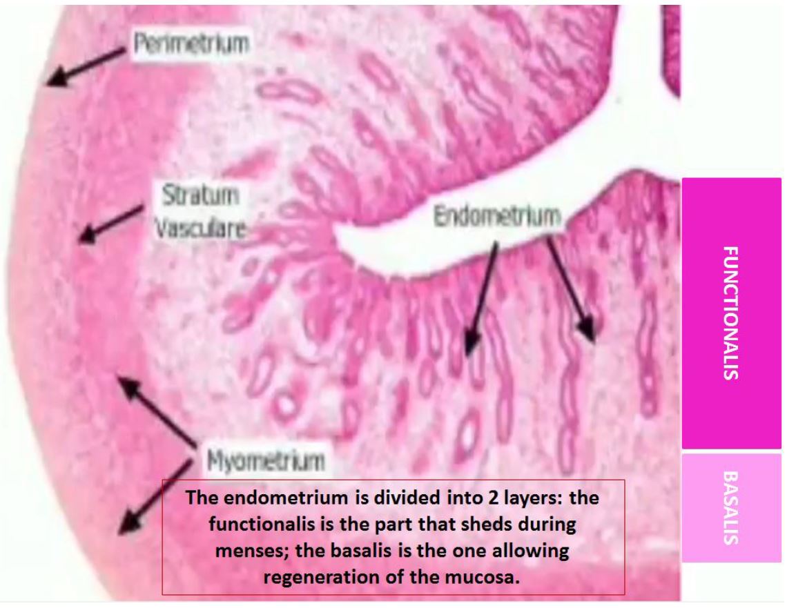
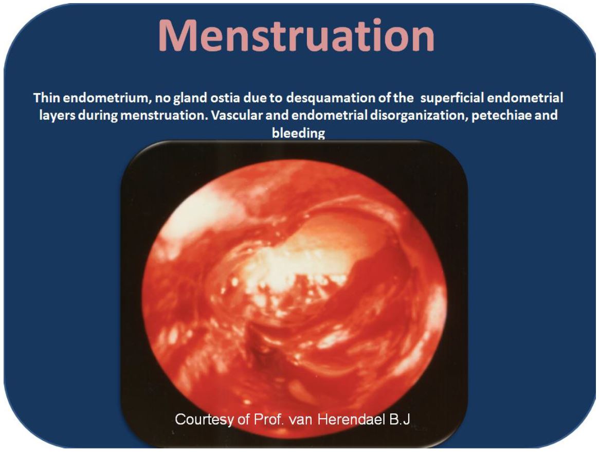
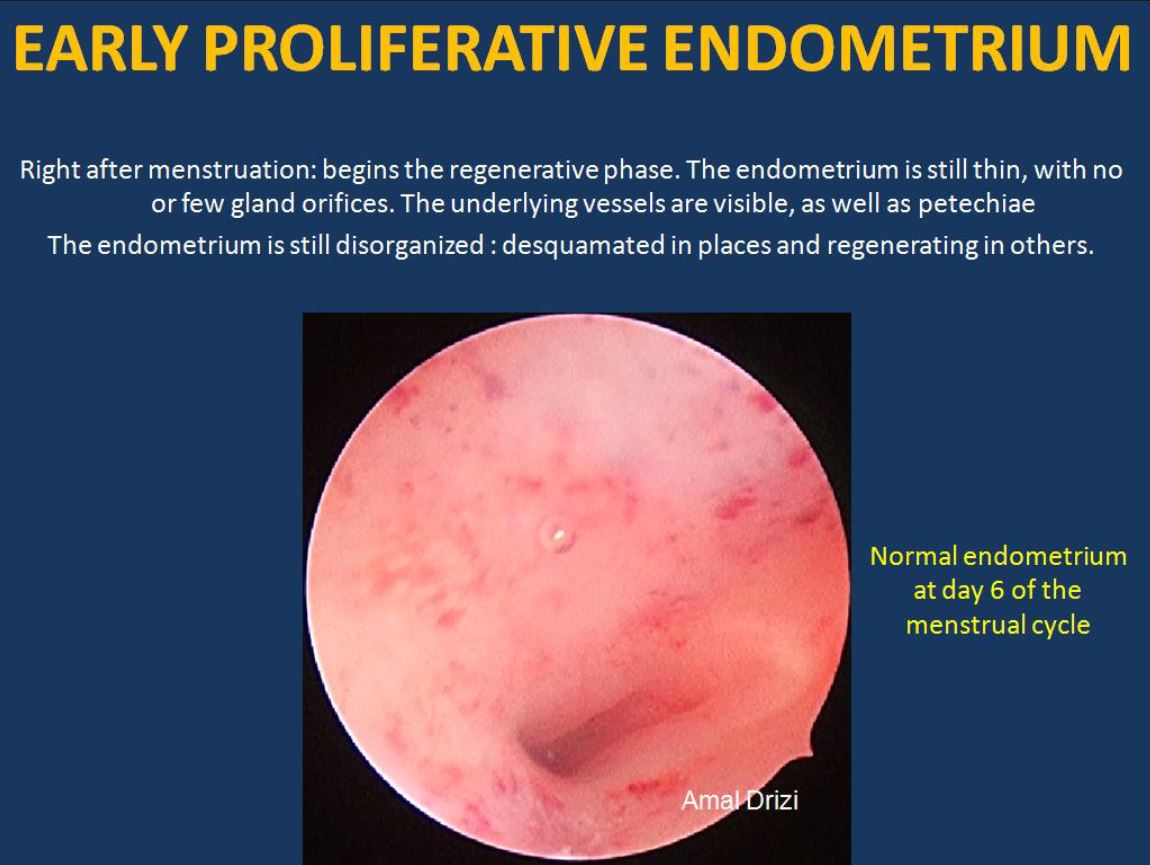
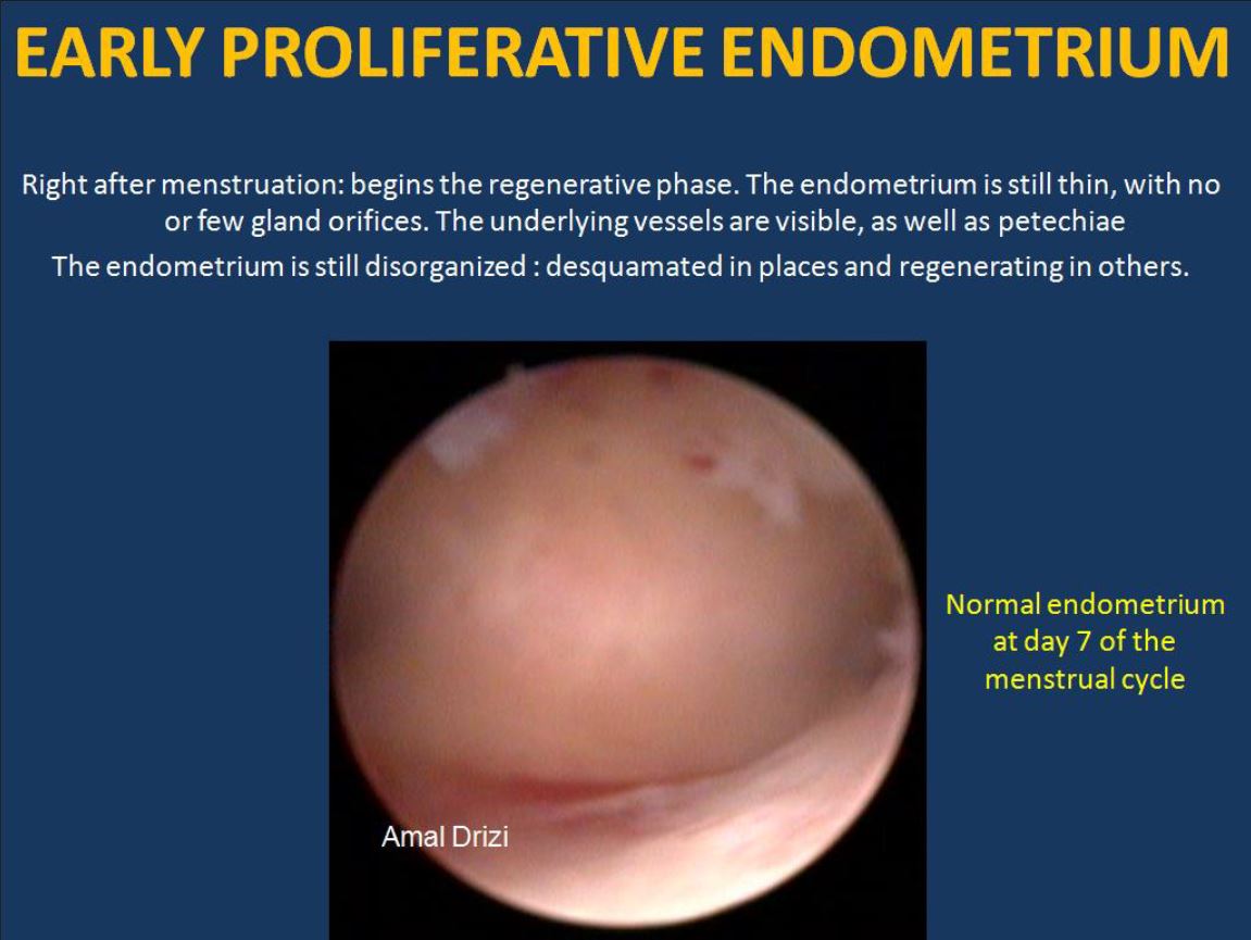
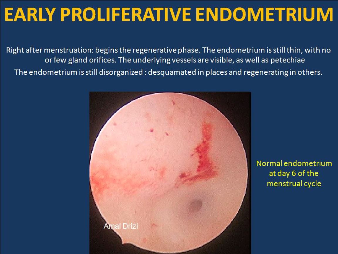
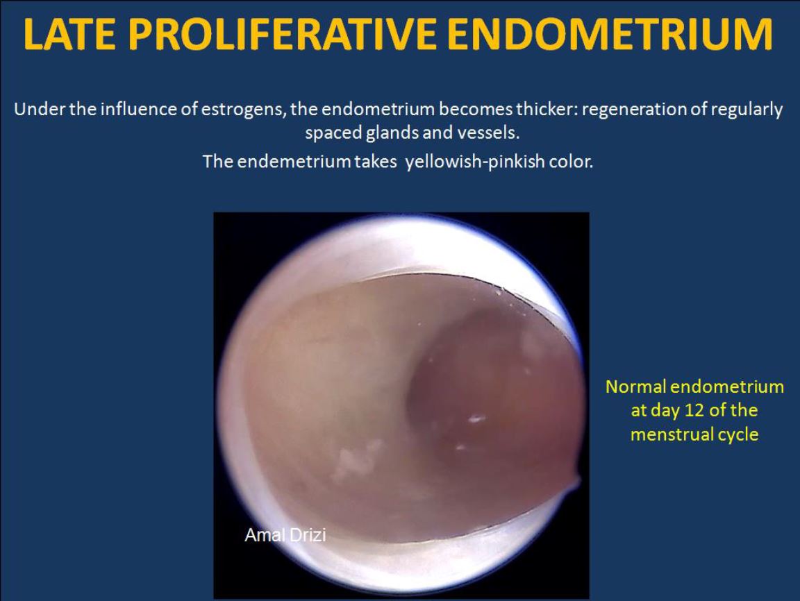
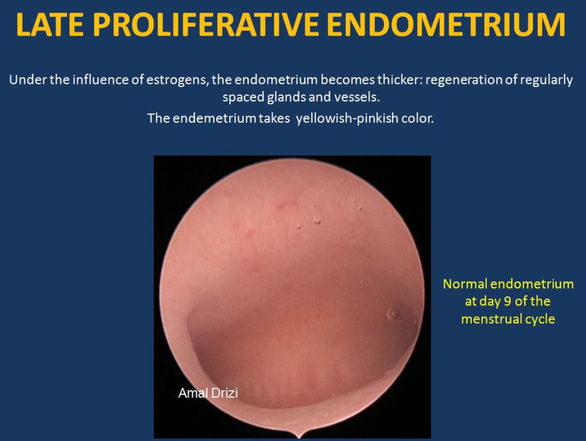
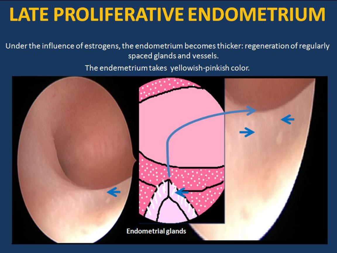
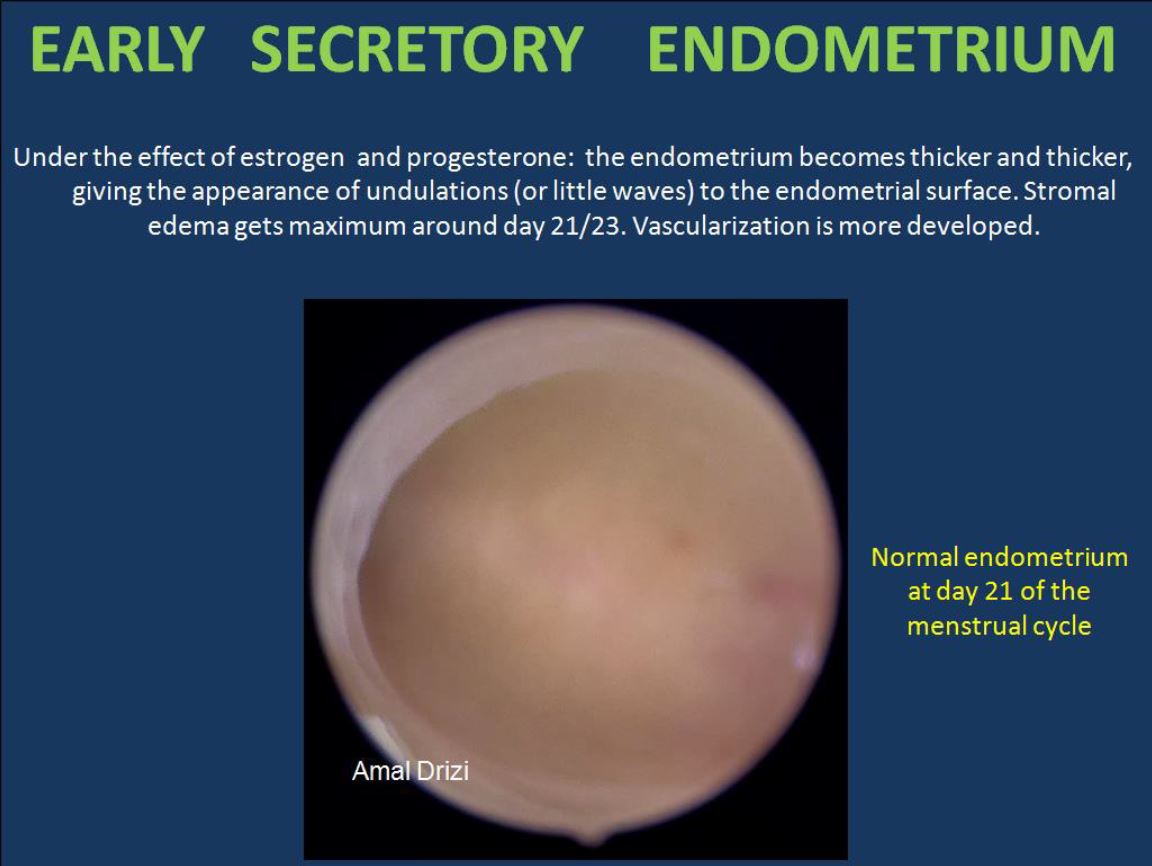
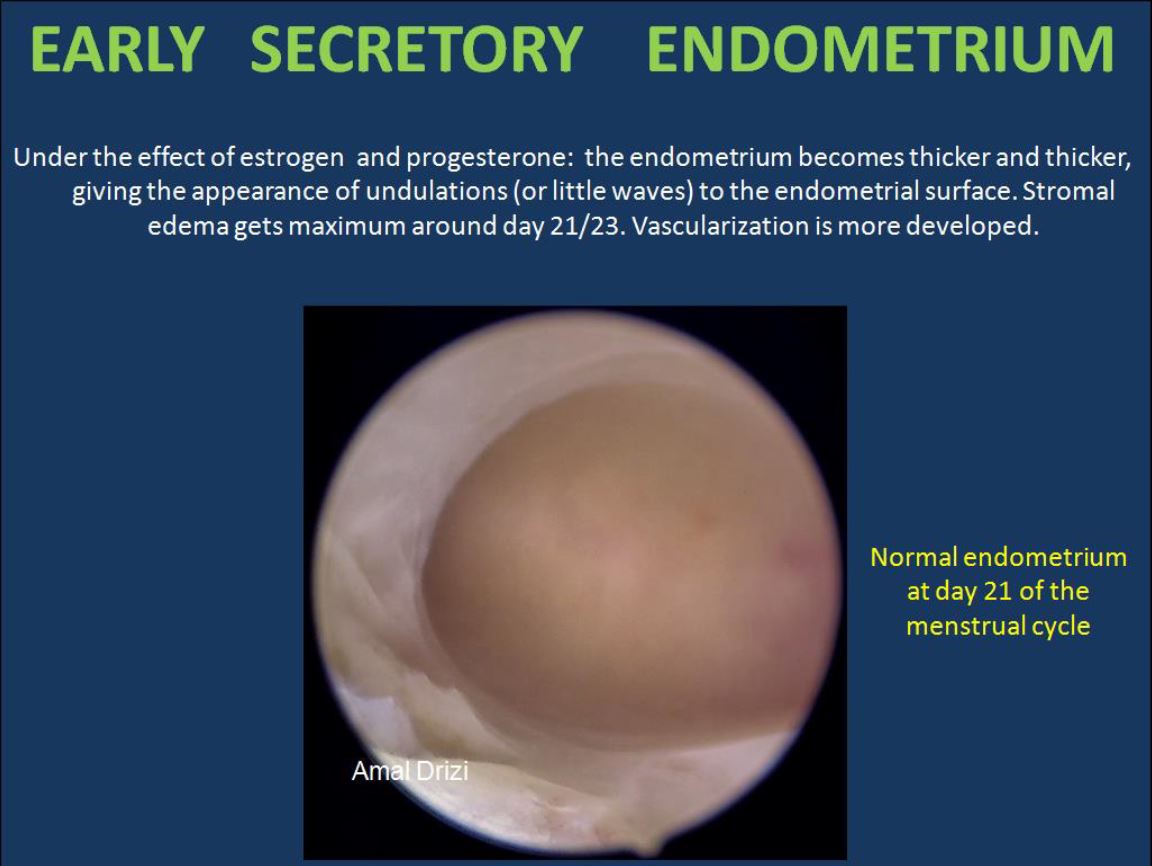
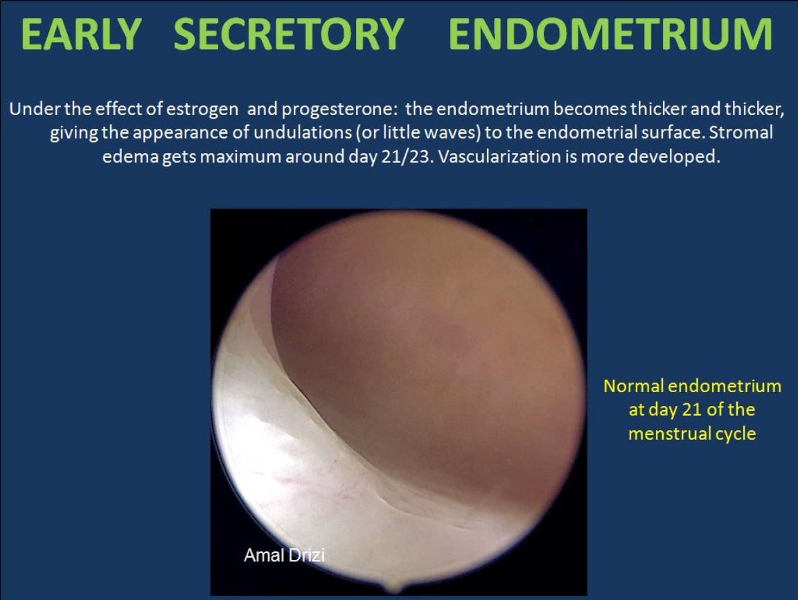
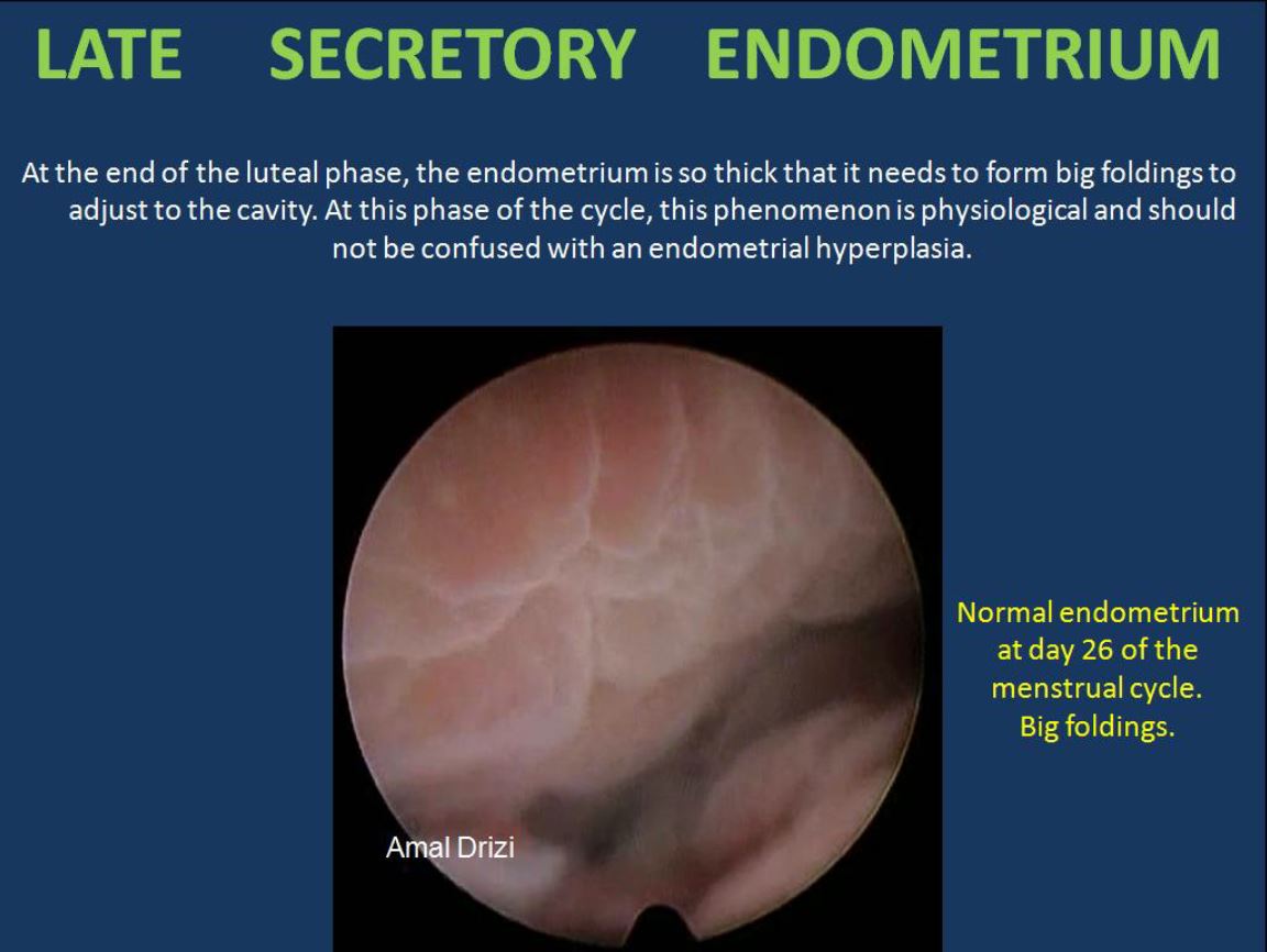
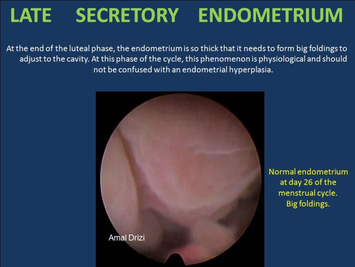
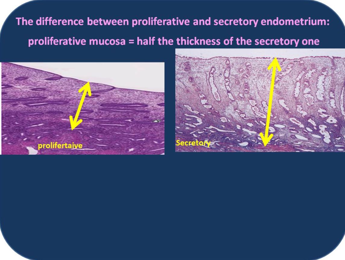
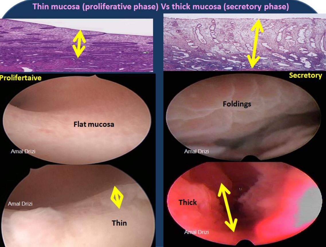
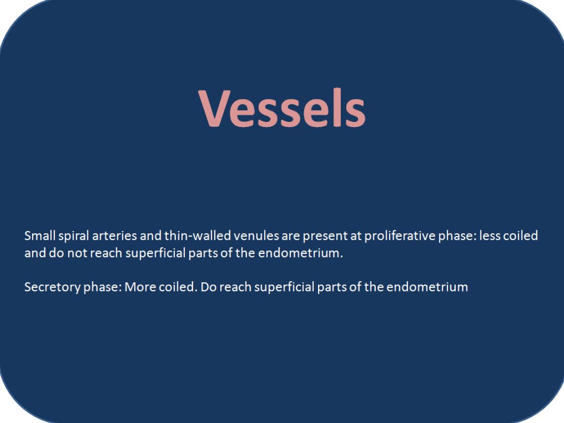
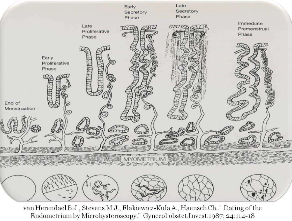
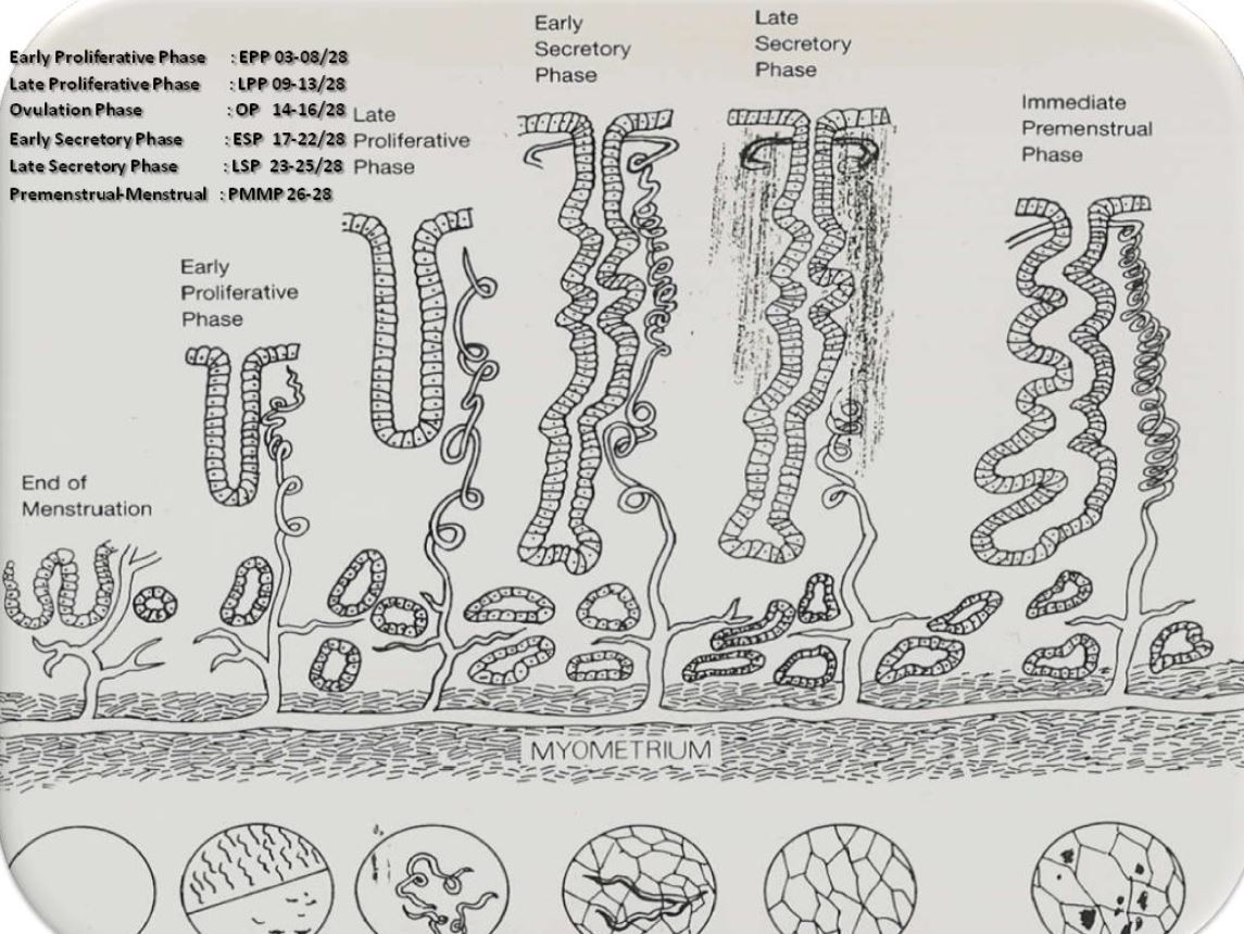
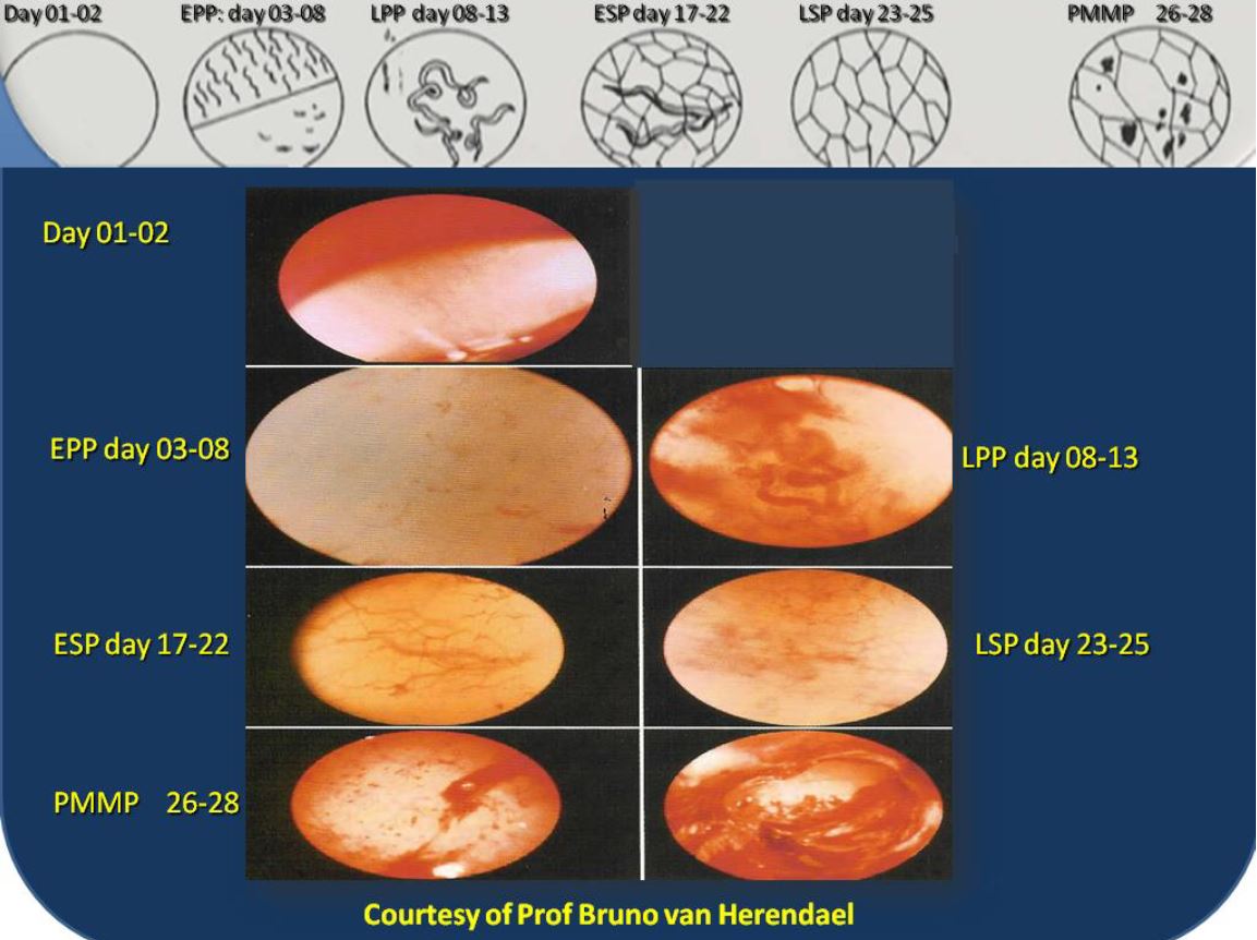
The description of the endometrium is based on histological bases to explain the different layers under the surface epithelium – visible with the diagnostic hysteroscope. These findings are then correlated with the visible features, through the hysteroscope in a non-contact mode. These findings are related to the morphologic and histologic changes allowing for the dating of the endometrium. The different specific phases of the endometrial cycle are described by pictures.
Discussion
The histological features of the Endometrium are correlated with the hysteroscopic aspect and can be a guide to screen the Endometrium during diagnostic procedures. However, the final diagnosis remains with the pathologist.

